Bioprinting – Made of Light
What if Serious Diseases Could Be Mimicked in Printed Organ Models?
Life itself is 3D, not 2D. Only structured, three-dimensional models can truly replicate the intricacies of living organisms.
Developing new medicines and treatments often involves extensive animal testing and clinical trials due to the complexity of replicating the 3D architecture of organs. Xolography offers a revolutionary way to print cells in defined 3D environments, enhancing the accuracy and efficacy of medical research.
No Stress while Printing
A stationary gel matrix is solidified in a rapid print - not applying any stress to material nor cells and maximizing cell viability.

Extra Soft Hydrogels
Printed materials may be exceptionally soft to resemble stiffness of almost all natural tissue structures.
No Sedimentation
The gelly hydrogel matrix embeds and nourishes the cells softly preventing uncontrolled sinking.
Status Quo
Bioprinting has focused on 2D methods, effective for skin but problematic for complex 3D organs. Layer-by-layer printing stresses cells and causes damage. Extrusion and DLP printers lack precision, while 2PP printers are too slow. What's missing is a mix of diverse materials, cell-friendly processing, and precise, cost-effective large-scale printing.
With Xolography
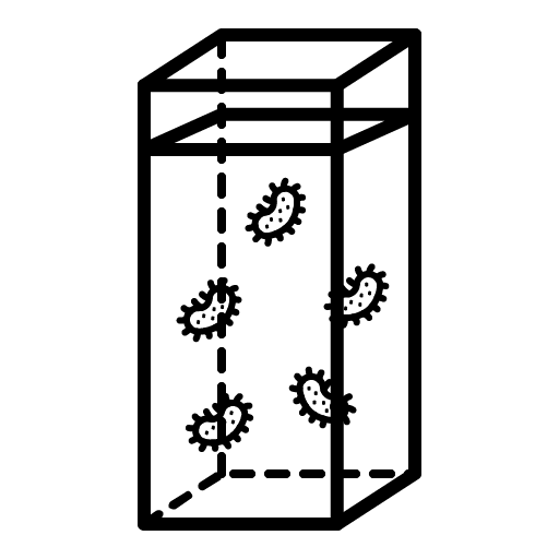
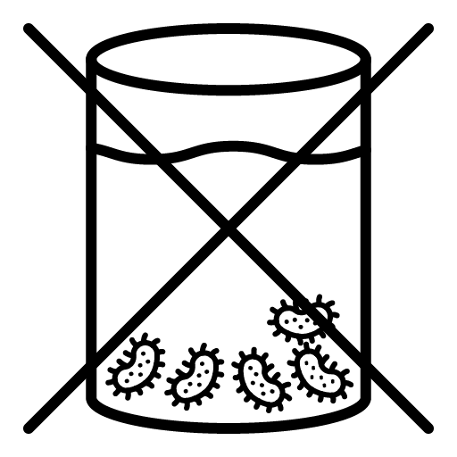
Xolography excels in producing large objects with seamless vascularization and gentle cell handling. Sealable cuvettes enable sterile models and cleaner prints than open systems. This comprehensive approach and its printing speed positions Xolography to drive the next breakthrough in Bioprinting.
How Xolography opens up new potentials in Bioprinting
Bioprinters create structures that mimic the 3D architecture of organs, aiming to provide the ideal environment for cells. The main challenge in bioprinting today is vascularization - forming a fine network of blood vessels for oxygen and nutrient supply deep into tissue. Current models are limited in size and complexity, resembling cell clusters rather than full organs.
However, with advanced Bioprinting, patient-derived cells could be used for personalized drug testing, and printed organ models could deepen our understanding of diseases. Xolography holds promise for producing tissue implants from human stem cells, potentially revolutionizing regenerative medicine.
The greatest potential lies in saving time. Faster, more targeted studies accelerate the journey from hypothesis to therapy, ultimately saving lives.